Pearly Penile Papules Treated with the Erbium:YAG Laser
Matilda Bylaite and Thomas Ruzicka
Department of Dermatology, Heinrich-Heine-University Duesseldorf, Duesseldorf, Germany
History
A 19-year-old man was referred with an 8-year history of persistent papular lesions around the coronal rim of his glans penis. The last few years he has been treated with various topical wart preparations, including podophyllin without any therapeutic success. The patient was anxious to be effectively treated, since he was very embarrassed by the penile lesions and his female partner’s reaction. His history included circumcision, bronchial asthma in childhood and allergic rhinitis. He had one sexual partner, was using condoms as contraception, and denied any history of sexually transmitted diseases. Clinical Findings
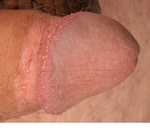
A genital examination revealed smooth, skin-colored, dome-shaped ample discrete papules 1 to 4 mm in size, distributed in three to five rows circumferentially along the coronal sulcus of the glans penis (Figs. 1 and 2). The patient was circumcised. There was no lymphadenopathy.
| Fig. 1. Pearly penile papules (PPP) are circumferentially distributed on the corona of the glans penis. |
 |

  |
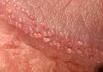
| Fig. 2. Close-up view of penile angiofibromas (PPP). |
 |
|
Histopathology
A biopsy was not performed. Examination and Laboratory Findings
Routine laboratory test were within normal limits. Human papilloma virus (HPV) test in material obtained by scrap of the corona penis was negative. Diagnosis
A clinical diagnosis of pearly penile papules was established. Therapy and Course
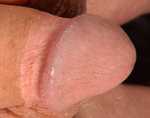
The patient was treated with the Erbium:YAG laser in two steps. After the first treatment, our patient was instructed to apply a topical povidone-iodine cream as antiseptic to the treated areas for the following 2 weeks. The wound healing was not compromised. Follow-up 1 month later demonstrated excellent cosmetic results (Fig. 3) with only a few discrete papules left. The treated lesions had resolved without scarring or post-inflammatory pigmentary changes. The patient was very satisfied with the results and requested a complete removal of the lesions.
| Fig. 3. Clinical regression of penile papules after one Erbium:YAG laser treatment. |
 |
|
Discussion
Pearly penile papules (PPP) are smooth, flesh-colored papular lesions, frequently found in men after puberty, and distributed circumferentially on the corona and sulcus of the glans penis. PPP are asymptomatic acral angiofibromas (1) that represent a physiologic variant of the normal penis and do not require treatment. However, the appearance of the lesions is often a reason for anxiety of both sexual partners and may cause a significant psychological and physiological distress. Moreover, PPP are most often mistaken for viral warts or sebaceous hyperplasia and usually unsuccessfully treated in clinics for sexually transmitted diseases. The incidence rate of PPP has been reported to be approximately 35% around 20 to 30 years of age, with declined rates below 15% after 40 years (2-4). The higher prevalence has been described in black and circumcised men (2). Previous reports suggested that HPV could play a causative role in the genesis of PPP (5); however, recently it has been denied since the lesions of PPP did not contain HPV DNA sequences (6, 7). Moreover, histologically PPP lack the characteristic morphologic features of HPV infection, showing a core of normal connective tissue covered by a centrally thin and peripherally acanthotic epidermis, over a vascular proliferation surrounded by a lymphocytic infiltrate and dense connective tissue (1). Interestingly, PPP share histologic features with other acral angiofibromas, such as subungual and periungual fibromas, fibrous papule of the face, acquired acral fibrokeratoma, and oral fibroma (1). PPP is a variation of the normal male anatomy. The reason to recognize these papules is to reassure worried men of their benign origin and to avoid unnecessary invasive treatments to remove them. However, many patients request treatment. Various treatment modalities, including podophyllin, circumcision, curettage, electrodesiccation, cryotherapy and carbon dioxide laser were employed with varying cosmetic success (8-10). Usually, the lesions are removed under local anesthesia after one or two treatments. Pigmentary changes and scarring might occur after treatment. We describe a male patient with pearly penile papules who was successfully treated with the Erbium:YAG laser. Familiarity with such physiological variants resembling dermatoses helps to avoid anxiety of the patients, providing them an adequate treatment. References
1. Ackerman, A.B., Kornberg, R. Pearly penile papules: acral angiofibromas. Arch Dermatol 1973, 108: 673-5. 2. Glicksman, J.M., Freeman, R.G. Pearly penile papules: a statistical study of incidence. Arch Dermatol 1966, 93: 56-9. 3. Rehbein, H.M. Pearly penile papules: incidence. Cutis 1977, 19: 54-7. 4. Rufli, T., Eichenberger, P., Heer, K. Papillomatosis coronae glandis. Frequency of occurrence and clinical picture. Schweiz Med Wochenschr 1978, 18: 229-31. 5. Mayeaux, E.J. Jr., Harper, M.B., Barksdale, W., Pope, J.B. Noncervical papillomavirus genital infectious. Am Fam Physician 1995, 52: 1137-50. 6. Ferenczy, A., Richart, R.M., Wright, T.C. Pearly penile papules: absence of human papillomavirus DNA by the polymerase chain reaction. Obstet Gynecol 1991, 78: 118-22. 7. Hogewoning, C.J., Bleeker, M.C., van den Brule, A.J. et al. Pearly penile papules: still no reason for uneasiness. J Am Acad Dermatol 2003, 49: 50-4. 8. Magid, M., Garden, J.M. Pearly penile papules: treatment with the carbon dioxide laser. J Dermatol Surg Oncol 1989, 15: 552-4. 9. Ocampo-Candiani, J., Cueva-Rodriguez, J.A. Cryosurgical treatment of pearly penile papules. J Am Acad Dermatol 1996, 35: 486-7. 10. Porter, W.M., Bunker, C.B. Treatment of pearly penile papules with cryotherapy. Br J Dermatol 2000, 142: 847. |