The Therapeutic Approach of Demodex Folliculitis Conglobata
Matilda Bylaite and Thomas Ruzicka
Department of Dermatology, Heinrich-Heine-University Duesseldorf, Duesseldorf, Germany
History
A 44-year-old woman presented with a 2-week history of severe exacerbation of acneiform facial lesions, strong headache and fever, which developed during her spring holidays in Tunis. Her medical history included mild acne for many years, struma, pollinosis and allergy (generalized exanthem) to penicillin. Treatment with oral ciprofloxacin and intravenous corticosteroid injections failed to relieve the skin manifestations. Clinical Findings
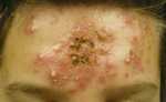
Physical examination revealed multiple confluent erythematous papules, pustules and infiltrated painful nodules, centrally covered with yellowish and hemorrhagic crust, and localized on an erythematous and edematous background of her forehead (Fig. 1). There was no evidence of telangiectases. Her temperature was 37.8º C and her regional lymph nodes were enlarged.
| Fig. 1. A 44-year-old woman present Demodex folliculitis conglobata on her forehead. |
 |

  |
Histopathology
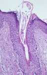
A skin biopsy specimen showed follicular hyperkeratosis with a mild perifollicular inflammatory infiltrate and Demodex mites in the dilated follicular infundibulum (Fig. 2).
| Fig. 2. Demodex folliculitis conglobata: Demodex mites are seen in the dilated follicular infundibulum (HE x100). |
 |
|
Examination and Laboratory Findings
Routine laboratory tests were within normal ranges. Bacterial and mycological cultures of skin swabs and pustules contents were negative. A large number of mites were seen in skin scrapings. Diagnosis
Based on clinical manifestation of folliculitis conglobata and detection of Demodex mites in the biopsy specimen and skin scrapings, a diagnosis of Demodex folliculitis conglobata was established. Therapy and Course
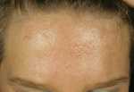
The patient was treated with oral 250 mg metronidazole 3x a day for two weeks. Additionally, topical 2% metronidazole gel and 0.15% lindane emulsion was continuously applied for two months till complete recovery was achieved (Fig. 3). After 3 weeks of therapy, the skin scrapings were negative for Demodex. Follow-up for next 12 months showed excellent control of the disease.
| Fig. 3. Clearing of Demodex lesions after 2 months of combined therapy with oral and topical metronidazole and lindane lotion. |
 |
|
Discussion
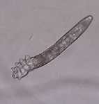
Demodex mites are common asymptomatic parasites of normal human skin. Demodex folliculorum, a 0.3 mm long transparent mite of the pilosebaceous follicles (Fig. 4), was first described in 1842 (1). Another species identified in humans is Demodex brevis that preferably resides in the sebaceous glands. The highest concentration of Demodex mites is found in the skin of the face, particularly on the nose, cheeks, eyelids and forehead, where sebaceous glands are numerous (2-4). The prevalence of infestation with Demodex increases with age, and in adults it is about 90-100% (3, 4). In children, Demodex are rare and almost exceptional in neonates (5); however, immunosuppression, such as lymphoproliferative disorders or AIDS, might facilitate the infestation (5, 6). Demodex has been implicated in development of various clinical disorders such as follicular pityriasis, pustular folliculitis, facial abscesses, rosacea-like demodicosis, granulomatous rosacea, perioral dermatitis, blepharitis, papulopustular scalp eruption, and also in facial lesions of immunosuppressed patients (2, 3, 7, 8). However, the pathogenic role of Demodex mites remains unclear and controversial (2). Since Demodex is found in healthy individuals, the mite density is the most important factor. Demodex has been suggested to play a pathogenic role in manifestation or exacerbation of facial conditions when the density is 5 or more mites/mc2. The diagnosis of Demodex infestation can be easily confirmed by direct microscopic examination of skin scrapings, expressed follicular contents, adhesive tape, cyanoacrylate glue (skin surface biopsy) and skin biopsy specimens. Interestingly, the number of mites varies greatly with the site examined and the method employed (3, 9). A classical skin biopsy shows a perifollicular infiltrate with the presence of numerous Demodex in the dilated ostium of hyperkeratotic follicles (10). One could also observe dilated vessels and extrafollicular granulomatous reactions to Demodex in the dermis. Recently, a noninvasive sampling method, a so-called standardized skin surface biopsy (SSSB), has been successfully employed (9). However, SSSB can fail to reflect the complete biotope of the Demodex, which can be much higher, since it collects only the superficial part of the skin (11). The differential diagnosis includes acne conglobata, rosacea conglobata, bromoderma, pyogenic infection, acneiform drug reactions, deep dermatophyte infection, trichophytosis, eosinophilic folliculitis, Demodex folliculitis and Demodex abscesses. Several therapeutic modalities have been reported to be effective in eradication of Demodex. The use of topical and systemic acaricidal agents, such as permethrin, lindane, crotamiton, malathion and benzyl benzoate, as well as oral ivermectin may lead to rapid and complete recovery (3). Alternatives are oral erythromycin, and topical or systemic metronidazole (3, 5, 6, 8). We describe a 44-year-old woman who developed severe facial lesions due to Demodex infestation and was successfully treated with oral metronidazole in conjunction with topical metronidazole gel and lindane emulsion. Further studies are necessary to better underst and the pathogenic role of Demodex in various skin manifestations, and provide evidence-based support in a large group of patients for this therapeutic approach.
| Fig. 4. Demodex folliculorum. |
 |
|
References
1. Simon, G. Die Haarsackmilbe. Medizinische Zeitung vom Vereine für Heilkunde in Preussen. 1842, No. 9.
2. Buns, D.A. Follicle mites and their role in disease. Clin Exp Dermatol 1992, 17: 152-5.
3. Forton, F., Seys, B., Marchal, J.L., Song, M. Demodex folliculorum and topical treatment: acaricidal action evaluated by standardized skin surface biopsy. Br J Dermatol 1998, 138: 461-6.
4. Bonnar, E., Eustace, P., Powell, F.C. The Demodex mite population in rosacea. J Am Acad Dermatol 1993, 28: 443-8.
5. Castanet, J., Monpoux, F., Mariani, R., Ortonne, J.P., Lacour, J.P. Demodicosis in a immunodeficient child. Pediatr Dermatol 1997, 14: 219-20.
6. Barrio, J., Lecona, M., Hernanz, J.M. et al. Rosacea-like demodicosis in a HIV-positive child. Dermatology 1996, 192: 143-5.
7. Ayers, S. Demodex folliculorum as a pathogen. Cutis 1986, 37: 441.
8. Schaller, M., Sander, C.A., Plewig, G. Demodex abscesses: clinical and therapeutic challenges. J Am Acad Dermatol 2003, 49: S272-4.
9. Forton, F., Seys, B. Density of Demodex follicularum in rosacea: a case-control study using standardized skin - surface biopsy. Br J Dermatol 1993, 128: 650-9.
10. Aylesworth, R., Vance, J.C. Demodex folliculorum and Demodex brevis in cutaneous biopsies. J Am Acad Dermatol 1982, 7: 583-9.
11. Forton, F., Song, M. Limitations of standardized skin surface biopsy in measurement of the density of Demodex follicularum. A case report. Br J Dermatol 1998, 139: 697-700. |