Treatment of Severe Atopic Eczema
Matilda Bylaite and Thomas Ruzicka
Department of Dermatology, Heinrich-Heine-University Duesseldorf, Duesseldorf, Germany
History
A 40-year-old man presented to our department with a 2-month history of severe eczema, intensive pruritus and sleep disturbance. His medical history revealed atopic eczema and allergic rhinitis diagnosed early in childhood. Previous atopy patch tests with common aero-allergens disclosed sensitization against most common pollen, house-dust mites and animal dander. Since his last hospitalization 7 years ago, the disease has been satisfactorily controlled with irregular use of emollients, topical corticosteroids, antihistamines and, rarely, UV-light therapy. Recently, emotional stress led to a generalized and severe flare of atopic eczema. There was also a family history of atopy, as the patient’s sister had an atopic eczema and his uncle suffered from asthma. Clinical Findings
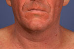
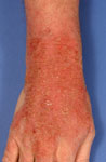
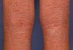
Physical examination revealed ill-defined, symmetrical, edematous and erythematous patches and plaques with fine scaling and yellowish crusting. The eczematous lesions were particularly distributed over the face and skin flexures, including eyelids, neck, flexural fossae, wrists and dorsal aspects of the hands and feet (Figs. 1 and 2). Less severe changes were noticed on the trunk and thighs. The skin was diffusely dry with numerous excoriations and pronounced lichenification in flexures (Fig. 3). There was no obvious lympadenopathy. White dermographism, as well as characteristic atopic stigmata, such as cheilitis, periorbital pigmentation, Dennie-Morgan fold and hyperlinear palms were evident.
| Fig. 1. Severe atopic eczema in the face: erythematous infiltration, scaling, crusting and excoriations. Similar changes were observed in skin flexures, and less severe changes on the trunk (before). |
 |

  |
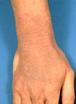
| Fig. 2. Severe atopic eczema of dorsal aspect of the hand before treatment. |
 |
|
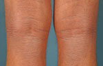
| Fig. 3. Note infiltration, lichenification and numerous excoriations in popliteae fossae before treatment. |
 |
|
Histopathology
Skin biopsy was not performed. Previous histopathological examination of a biopsy specimen taken in 1997 had shown chronic spongiotic dermatitis, thus supporting the diagnosis of atopic eczema. Examination and Laboratory Findings
Laboratory tests disclosed increased CRP (10.2 mg/L, normal 1.5-5.0 mg/L), mild eosinophilia, markedly elevated total IgE (>5000 kU/l, normal <20 kU/l) and eosinophil cationic protein (>2000 µg/l, normal <18 µg/l) levels in serum. Radioallergosorbent test (RAST) revealed significantly elevated allergen-specific IgE levels against house-dust mites (CAP-class 6), Aspergillius fumigatus (CAP-class 3) and Pityrosporum orbiculare (CAP-class 3). Blood cell count and other routine serum biochemistry findings were within normal ranges. Diagnosis
Based on his previous medical history, clinical and laboratory findings, a diagnosis of chronic generalized atopic eczema in association with allergic rhinitis was established. Therapy and Course
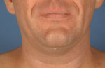
Therapy was initiated with intensive skin care, including hydration (oil baths) followed by application of emollients and wet-wrap bandages as a basic daily treatment. The patient was reluctant to take any systemic medication except for sedating antihistamines for intensive pruritus and better sleep. After 2 days of intensive skin care, additionally the potent topical steroid Dermoxin® (clobetasol-17-propionate) ointment twice daily was applied over lesional skin for 2 days. In the next 5 days, only topical tacrolimus 0.1% ointment twice daily was initiated. Acetylsalicylic acid 500 mg was prescribed 30 min before application of tacrolimus ointment to reduce local symptoms, such as transient pruritus and burning sensation. This treatment resulted in a significant improvement of the patient’s condition within 1 week. After 10 days of therapy, he was discharged with dramatically improved eczematous lesions (Figs. 4, 5 and 6). However, moderate lichenification in skin flexures remained. Skin care, including hydratation and emollients, long-term topical tacrolimus 0.1% ointment twice daily and stress management techniques were suggested. The patient used environmental measures to reduce the impact of house-dust mites on his skin. One month later, our patient remained under excellent symptomatic control with topical tacrolimus. He showed no side effects due to tacrolimus 0.1% ointment, and his quality of life was markedly improved.
| Fig. 4. After 1 week of treatment. |
 |
|
| Fig. 5. Severe atopic eczema of dorsal aspect of the hand after 1 week of treatment. |
 |
|
| Fig. 6. Popliteae fossae after 1 week of treatment. |
 |
|
Discussion
Atopic eczema (AE) is a common, intensively pruritic, chronically relapsing, inflammatory skin disease, significantly affecting patient’s health and quality of life (1, 2). Generally, over 50% of patients manifest eczematous lesions by the first year and over 90% before 5 years of age (3). Rarely, atopic eczema has an adult onset, which is often severe. The prevalence of AE is about 15-20% in the general population; however, recent epidemiological reports indicate its increasing frequency over the past decades (4). Furthermore, 40-60% of patients with AE have respiratory allergies (5). The pathogenesis of AE is multifactorial. The disease has a genetic basis, and its clinical expression is modified by numerous exogenous and endogenous factors (5). The humoral and cellular immunity is known to be disturbed, thus a relative TH1/TH2 imbalance, overproduction of cytokines and total IgE results in hypersensitivity to environmental factors that normally are not allergens (3). Moreover, it has been shown that increased IgE receptors on the surface of Langerhans cells in atopic patients bind inhalant allergens in the epidermis and evoke a delayed-type contact hypersensitivity reaction, which is of diagnostic use in atopy patch tests (5). The clinical picture of AE in adults is variable. Usually, the disease is moderate with a relapsing and remitting course, affecting any region of the body, predominantly skin flexures. However, 2% of patients run a more severe course with larger areas of skin involvement (6). Face and neck dermatitis, as well as chronic hand dermatitis are often of major concern due to their visibility and complicated therapy. Even though Hanifin and Rajka criteria are the most widely used to describe AE (1), recently, Bos et al. proposed the millennium criteria, emphasizing the presence of allergen-specific IgE in serum (7). The therapeutic management of AE should consider its chronic and relapsing nature and include both preventive and therapeutic measures. Although a broad range of therapeutic options that provide a symptomatic relief is available, interaction with itch-scratch cycle and safe and long-term disease control remain challenging. Daily skin care, emollients, preventive life style and environmental measures are critical; however, in many patients they are not sufficiently effective to prevent disease exacerbation. The traditional treatment includes topical corticosteroids, antihistamines and antibiotics. Corticosteroids are still valuable for acute flares in very severe or refractory AE. Unfortunately, a long-term use of corticosteroids or use on the face, neck and other intertriginous areas is limited for their well-known side effects. Tacrolimus ointment, a novel steroid-sparing topical immunomodulator, has been shown to be as effective as mild to potent corticosteroids in the short-term management of AE symptoms, and safe in intermittent long-term treatment without the risk of corticosteroid-associated side effects (6, 8, 9). Currently, topical tacrolimus was also successfully employed in the treatment of a variety of other inflammatory dermatoses, such as psoriasis, acrodermatitis continua, lichen planus, lichen sclerosus, pyoderma gangrenosum, leg ulcers and steroid-induced rosacea (10). In summary, we describe a patient with severe atopic eczema whose symptoms were successfully controlled with topical agents. Evidently, topical treatment, including intensive skin care and repeated applications of potent steroids for two days, followed by only tacrolimus ointment application for five days, was very effective in our patient. A rapid control of symptoms and the inflammatory process with topical regimen led to a dramatic improvement of his skin condition. His quality of life markedly improved. Thus, we consider prolonged therapy with topical tacrolimus, in some cases in short-term combination with topical corticosteroids, and basic skin care to be of significant benefit in treating AE. References
1. Hanifin, J.M., Rajka, G. Diagnostic features of atopic dermatitis. Acta Derm Venereol 1980, 92: 44-7.
2. Finlay, A.Y. Quality of life in atopic dermatitis. J Am Acad Dermatol 2001, 45: 64-6.
3. Braun-Falco, O., Plewig, G., Wolff, H.H., Burgdorf, W.H.C. Atopic dermatitis. In: Dermatology: second, completely revised edition. Springer-Verlag: Berlin 2000, 499-509.
4. Diepgen, T.L. Epidemiology of atopic dermatitis. In: Tacrolimus Ointment: A Topical Immunomodulator for Atopic Dermatitis. T. Ruzicka, S. Reitamo (Eds.). Springer-Verlag: Berlin 2004, 3-19.
5. Ruzicka, T. Atopic eczema between rationality and irrationality. Arch Dermatol 1998, 134: 1462-9.
6. Reitamo, S. Clinical experience in adults. In: T. Ruzicka, S. Reitamo (Ed.). Tacrolimus ointment: A Topical Immunomodulator for Atopic Dermatitis. Springer-Verlag: Berlin 2004, 129-60.
7. Bos, J.D., Van Leent, J.M., Sillevis Smitt, J.H. The millennium criteria for the diagnosis of atopic dermatitis. Exp Dermatol 1998, 7: 132-8.
8. Ruzicka, T., Bieber, T., Schopf, E. et al. A short-term trial of tacrolimus ointment for atopic dermatitis. N Engl J Med 1997, 337: 816-21.
9. Reitamo, S., Wollenberg, A., Schopf, E. et al. Safety and efficacy of 1 year of tacrolimus ointment monotherapy in adults with atopic dermatitis. Arch Dermatol 2000, 136: 999-1006.
10. Assmann, T., Ruzicka, T. Other potential dermatological indications for tacrolimus. In: Tacrolimus Ointment: A Topical Immunomodulator for Atopic Dermatitis. T. Ruzicka, S. Reitamo (Eds.). Springer-Verlag: Berlin 2004, 129-60. |