Acquired Unilateral Nevoid Telangiectasia Syndrome
Matilda Bylaite and Thomas Ruzicka
Department of Dermatology, Heinrich-Heine-University Duesseldorf, Duesseldorf, Germany
History
A 35-year-old, 23-weeks pregnant primigravida woman was referred to our outpatient clinic with a 4-month history of an asymptomatic, persistent, erythematous eruption on the right side of her upper chest wall, shoulder and medial aspects of her right arm. Her pregnancy had been unremarkable except for the development of a flushing eruption, intensified by emotions. She was otherwise healthy and had no history of liver disease, alcohol abuse or oral contraceptives use. No family member had similar lesions. Interestingly, the patient used to notice a slight transient flushing on the right side of her upper chest wall during puberty after alcohol intake. Clinical Findings
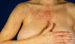
Physical examination revealed a macular network of numerous, grouped, erythematous fine telangiectatic vessels that blanched on pressure. The telangiectases were distributed in an unilateral, irregular dermatomal pattern (C3-C8), involving the right side of her upper chest wall, right shoulder and the medial aspect of the arm, extending along the volar aspect of her I-III fingers (Figs. 1 and 2). The mucous membranes were not involved. The Rumpel-Leede test was negative. There was no hepatosplenomegaly.
| Fig. 1. Unilateral nevoid telangiectasia syndrome (UNTS). A 35-year-old 23-week pregnant woman showing unilateral distribution of telangiectases. |
 |

  |
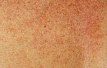
| Fig. 2. Unilateral nevoid telangiectasia syndrome (UNTS). Multiple fine telangiectases on the right aspect of the upper chest wall. |
 |
|
Histopathology
A skin biopsy was not performed. Examination and Laboratory Findings
Routine laboratory tests including complete blood cell count, blood glucose, liver and renal function tests, VDRL were within normal limits or negative. Studies of serum estrogen level were not performed. Diagnosis
A diagnosis of an acquired unilateral nevoid telangiectasia syndrome was made based on the history and the strictly unilateral arrangement of telangiectases in a pregnant woman. Therapy and Course
The telangiectases regressed spontaneously within one month after the birth of her healthy child. Three years later, physical examination demonstrated a completely normal skin without residual findings of telangiectases. Discussion
Unilateral nevoid telangiectasia syndrome (UNTS) is a rarely reported disorder, characterized by multiple telangiectatic lesions unilaterally distributed in certain dermatomes. The syndrome was first described in 1922 by Zeissler (1), and classified as primary telangiectases by Anderton and Smith in 1975 (2). Thereafter, Wilkin reviewed UNTS as a congenital and an acquired type (3). The acquired type is commonly associated with physiological or pathological hyperestrogenemic conditions, and divided into an estrogen-related group (puberty, pregnancy, hormonal therapy) and alcohol/hepatic-related group with or without systemic involvement (2). The pathogenesis of strictly unilateral development of telangiectases is still unknown. It has been proposed that UNTS may be a latent congenital estrogen-sensitive vascular nevus (4) that becomes evident due to idiosyncratic dermatomal sensitivity (5) to elevated serum estrogen levels or its metabolic products, and/or proliferation of both estrogen and progesterone receptors (6, 7), resulting in capillary dilatation. However, this theory does not suit all cases since an acquired UNTS was reported in a healthy man with a normal hormonal state and no history of alcohol abuse, underlying pathology or physiological condition (8). The role of epidermal angiogenic factor and prostaglandins has been also discussed (9). Recently, Kreft et al. presented microcirculatory examinations that showed pathophysiological defects at the vascular level even in subclinical cases of UNTS (10). UNTS usually presents as asymptomatic, numerous, small macular or raised telangiectatic lesions, predominantly affecting the face, neck, shoulder-arm region and thorax. Commonly, the eruption is found in the C3-T1 dermatomes (2). During pregnancy, menstrual cycle or progression of hepatic disorder the lesions usually increase in number, size and intensity of erythema. Histopathology of UNTS shows dilated superficial dermal vessels with no sign of endothelial cell proliferation in the upper and middle dermis. Since UNTS has usually only temporal cosmetic importance, destructive therapeutic procedures are not required. In cases associated with pregnancy, the telangiectatic lesions commonly fade within few weeks after delivery and completely regress soon after. In the hepatic-related group, the clinical evidence of UNTS is related to the severity of an underlying disease. We report a case of an acquired unilateral nevoid telangiectasia syndrome associated with pregnancy in a 35-year-old woman. Our case fulfills the criteria for classical acquired UNTS. The temporal presence of telangiectases during pregnancy suggests the obvious role of estrogen in a development of UNTS in our patient. Probably, UNTS is more common than reported. Further studies are still required to better understand the pathogenesis of this condition. References
1. Zeissler. Telangiectasia associated with syphilis and pregnancy. Arch Dermatol Syphil 1922, 5: 781.
2. Anderton, R.L., Smith, J.G. Unilateral nevoid telangiectasia with gastric involvement. Arch Dermatol 1975, 111: 617-21.
3. Wilkin, J.K. Unilateral nevoid telangiectasia. Three new cases and the role of estrogen. Arch Dermatol 1977, 113: 486-8.
4. Mirrer, E., Cipriano, A., McGuire, J. Unilateral nevoid telangiectasia. Arch Dermatol 1971, 103: 320-3.
5. Jucas, J.J., Rietschel, R.L., Lewis, C.W. Unilateral nevoid telangiectasia. Arch Dermatol 1979, 115: 359-60.
6. Uhlin, S.R., McCarty, K.S. Jr. Unilateral nevoid telangiectatic syndrome. The role of estrogen and progesterone receptors. Arch Dermatol 1983, 119: 226-8.
7. Karakas, M., Durdu, M., Sonmezoglu, S. et al. Unilateral nevoid telangiectasia. J Dermatol 2004, 31: 109-12.
8. Taskapan, O., Harmanyeri, Y., Sener, O., Aksu, A. Acquired unilateral nevoid telangiectasia syndrome. Acta Derm Venereol 1997, 77: 62-3.
9. Wolf, J.E., Harrison, R.G. Demonstration and characterization of an epidermal angiogenic factor. J Invest Dermatol 1973, 61: 130-41.
10. Kreft, B., Marsch, W.C., Wohlrab, J. Unilateral nevoid telangiectasia syndrome. Dermatology 2004, 209: 215-7. |