Acrodermatitis Enteropathica
Matilda Bylaite and Thomas Ruzicka
Department of Dermatology, Heinrich-Heine-University Duesseldorf, Duesseldorf, Germany
History
A 14-month-old, full-term, normal birth weight white boy presented with a 4-month history of psoriasiform plaques in perioral and perianal region, elbows and knees, associated with mild diarrhea, loss of appetite and irritability. The baby was weaned at 9 months age and 4 weeks later perioral lesions appeared. Within a short time, other parts of his body became affected. His mood changed and he became irritable. Previously, he had several upper respiratory tract infections. Initially, atopic dermatitis with impetigo was diagnosed and treated. Later, he was treated for epidermolysis bullosa simplex with sulfonamides, oral nystatin and local pimecrolimus cream in another hospital. Only slight improvement was noticed. Finally, the patient was referred to our hospital for evaluation and treatment. Clinical Findings
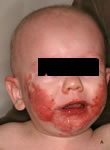
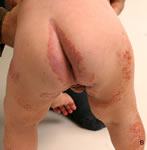
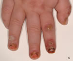
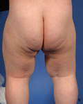
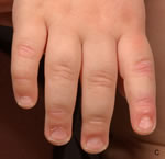
On physical examination, erythematous, confluent, sharply demarcated, symmetric, erosive, scaly and crusted plaques on his face were noted. The lesions, predominantly located in the perioral region extended over his cheeks and submental area ( Fig. 1 A). A few red, scaly papules were periorbital and around nostrils. Similar lesions were also found in the perigenital and perianal ( Fig. 1B) regions, and on the elbows and knees. Eczematous patches with papules were seen on lower limbs. Fingertips and toes were erythematous, oozing, with vesicles and pustules, painful fissures and paronychia ( Fig. 1C). Atrophic glossitis and superficial aphthous-like erosions were also found. There was diffuse alopecia and regional lymphadenopathy. The child was crying and irritated.
| Fig. 1A: Acrodermatitis enteropathica. Symmetric, confluent, eczematous-like eruption on the face and diffuse hair loss in a 14-month-old boy. |
 |

  |
| Fig. 1B: Acrodermatitis enteropathica. Similar lesions were found in the perianal, perigenital region and on his lower limbs. |
 |
|
| Fig. 1C: Acrodermatitis enteropathica. Fingertips show vesicles and pustules, painful fissures and paronychia |
 |
|
Histopathology
Histopathology was not performed. Examination and Laboratory Findings
Results of laboratory investigations revealed a markedly lowered zinc level (0.17 mg/l, normal 0.78-1.48 mg/l); copper serum level was 1.21 mg/l (0.82-1.39 mg/l). A potassium hydroxide examination was positive for Candida albicans. Normal cutaneous flora from skin lesions was found microscopically. Stool examination for pathogenic microorganisms was negative. Diagnosis
The diagnosis of acrodermatitis enteropathica was established. Therapy and Course
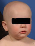
Oral zinc therapy 50 mg daily (5 mg/kg) was started. Within 2 days the irritability and diarrhea ceased, and after 2 weeks the skin lesions healed ( Fig. 2). The dose was reduced to 20 mg/day. At a follow-up 2 months later, the skin was free of lesions, and he continued to do well with blood zinc levels in the normal range.
| Fig. 2A: The lesions healed within 2 weeks after zinc replacement |
 |
|
| Fig. 2B: The lesions healed within 2 weeks after zinc replacement |
 |
|
| Fig. 2C: The lesions healed within 2 weeks after zinc replacement |
 |
|
Discussion
Acrodermatitis enteropathica (AE) is a rare autosomal recessive disorder of zinc absorption, presenting in infancy and characterized by a triad of circumorificial and acral dermatitis, alopecia and diarrhea. It starts within a few days or weeks of life, usually when the infant is weaned and placed on cow's milk (1). The latest studies have shown that low zinc levels in the mother's milk may cause an acquired zinc deficiency in full-term, breast-fed infants (2, 3). Similar clinical findings may also occur secondary to reduced dietary zinc intake, malabsorption (in celiac disease, bypass surgery, severe infantile diarrhea) (4, 5) or chronic alcoholism. Typically, dry, scaly, erythematous, sharply marginated eczematous patches and plaques present in the perioral and anogenital areas and later evolve into vesiculobullous, pustular, erosive and crusted lesions. Continuously, trunk, hands and feet are involved. Diffuse hair loss, diarrhea, paronychia and secondary infections are common. Depressed mood, irritation, photophobia and retardation of growth are frequently observed (5, 6). Low zinc levels in plasma and urinary excretion are the parameters to confirm the diagnosis. Histopathologic examination of skin biopsy specimens is nonspecific (2, 5). The list of differential diagnoses includes atopic dermatitis, seborrheic dermatitis, psoriasis, widespread mucocutaneous candidiasis, histiocytosis X, epidermolysis bullosa and acquired zinc deficiencies (4). As in our case, the initial suspicion of zinc deficiency is often missed. Before it was known that AE results from deficient zinc uptake, most patients died in childhood or suffered greatly. Oral zinc was introduced for therapy and to date remains the “gold standard”. The lesions heal spectacularly within 1 or 2 weeks, also diarrhea ceases and mood improves within 24 h. However, replacement should be lifelong. Here, we present an illustrative case of acrodermatitis enteropathica that has not been recognized and treated accordingly for a long time. The diagnosis of zinc deficiency should be considered whenever a patient presents with periorificial and acral lesions associated with alopecia and diarrhea, and adequate zinc supplement therapy should be started immediately. References
1. Danbolt, N., Closs, K. Acrodermatitis enteropathica. Acta Derm Venereol 1942, 23: 127-69.
2. Glover, M.T., Atherton, D.J. Transient zinc deficiency in two full-term breast-fed siblings associated with low maternal breast milk zinc concentration. Pediatr Dermatol 1988, 5: 10-3.
3. Roberts, L.J., Shadwick, C.F., Bergstressor, P.R. Zinc deficiency in two full-term breast-fed infants. J Am Acad Dermatol 1987, 16: 301-4.
4. Schachner, L.A., Hansen, R.C. Pediatric Dermatology. Churchill Livingstone Inc: New York 1988, p. 759.
5. Perafan-Riveros, C., Franca, L.F., Alves, A.C., Sanches, J.A. Jr. Acrodermatitis enteropathica: Case report and review of the literature. Pediatr Dermatol 2002, 19: 426-31.
6. VanWouwe, J.P. Clinical and laboratory diagnosis of acrodermatitis enteropathica. Eur J Pediatr 1989, 149: 2-8. |