Agminated Spitz Nevi within Nevus Spilus
Matilda Bylaite and Thomas Ruzicka
Department of Dermatology, Heinrich-Heine-University Duesseldorf, Duesseldorf, Germany
History
A 13-year-old red hair, fair-skinned boy was referred to our clinic for evaluation of a large congenital pigmented lesion on his left leg. The lesion had gradually enlarged, and over the last 4 years several reddish, itching nodules had developed within it. In addition, he had a 2-year history of myelodysplastic syndrome, controlled with ciclosporin A (40 mg/day). There was no family history of pigmented disorders. Clinical Findings
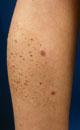
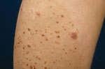
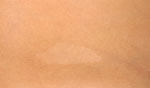
On examination, numerous, tiny, dark-brown macules and raised papules were scattered over a large, 12 x 12 cm in diameter, well-demarcated, light-brown patch on his left leg (Fig. 1). Multiple, grouped, 0.3-0.8 cm in size, dome-shaped, firm, light red-brown papules and nodules were also noticed within the patch (Fig. 2). A leaf-shaped white patch was found on his right hip (Fig. 3). Two ellipsoid, well-circumscribed, pale-brown patches were observed on the upper posterior part of his left leg and buttocks. Furthermore, the boy had multiple, discrete, flat tan-brown freckles over his central face.
| Fig. 1. Agminated Spitz nevi scattered over the nevus spilus. |
 |

  |
| Fig. 2. Close-up view of Spitz nevi within speckled lentiginous nevus. |
 |
|
| Fig. 3. Nevus depigmentosus. |
 |
|
Histopathology
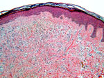
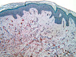
Histopathologic examination of a biopsy from a dark brown papule revealed dermal melanocytic nevus. Biopsy specimens, obtained from five different red-brown nodules showed hyperkeratosis and irregular acanthosis in the epidermis. Large, epithelioid melanocytes with abundant eosinophilic cytoplasm and large nuclei, and little or no melanin, were scattered in the dermis (Fig. 4). Immunohistochemical studies demonstrated a positive reaction of the tumor cells to S100 (Fig. 5) protein and negative to Melan-A. There were no signs of malignant changes in all obtained skin biopsies. These histological features were consistent with dermal Spitz nevi.
| Fig. 4. Spitz nevus. Large atypical melanocytes with abundant eosinophilic cytoplasm are scattered in the dermis (HE, x100). |
 |
|
| Fig. 5. Spitz nevus. Positive immunohistochemical reaction of tumor cells to S-100 protein (x100). |
 |
|
Examination and Laboratory Findings
Laboratory studies showed mild anemia and thrombocytopenia. Diagnosis
A diagnosis of agminated Spitz nevi arising within nevus spilus in association with nevus depigmentosus and café-au-lait patches in a patient with myelodysplastic syndrome was made. Therapy and Course
Serial surgical excisions were recommended for the patient. Sun protection and sunscreens, as well as regular 6-month follow-up for malignant changes has been advised. Discussion
Nevus spilus (NS, speckled lentiginous nevus, nevus on nevus, spotty nevus) is a relatively uncommon pigmented skin lesion, consisting of a large, light tan patch with numerous, circumscribed, dark brown macules or papules within a lesion. It was reported that NS occurs in less than 0.2% of all newborns and 1.2% of white school children (1). The nature of NS remains unclear. It is believed to be a localized defect in neural crest of melanoblasts. Genetic and environmental factors could also play a role (1, 2). Nevus spilus often presents at birth or during the first years of life as a hyperpigmented patch. In most cases, it takes several years to develop its characteristic picture with multiple dark speckles. It usually presents as a single lesion, but multiple lesions in a zosteriform distribution along a dermatome may be present (3). A biopsy obtained from the tan background shows histological features of lentigo simplex. The speckled areas show varying histological patterns that range from junctional, compound and dermal nevi to blue nevi or Spitz nevi (4, 5). NS can occur in association with cutaneous vascular malformations, such as nevus flammeus in cases of phacomatosis pigmentovascularis (6). In our patient, multiple agminated Spitz nevi slowly developed within the large nevus spilus. This condition is considered to be rare, as only few cases have been reported in the literature (7). Moreover, these lesions were associated with other pigmentary disorders, such as freckles, café-au-lait spots and nevus hipopigmentosus. Interestingly, 2 years after the first Spitz nevi started to appear, myelodysplastic syndrome was diagnosed. In our patient, histopathological examination confirmed the pigmentary lesions to be benign. However, recent reports of melanomas developing within nevus spilus have increased the awareness of these lesions as potential precursors of melanoma (5, 8, 9). Although the nature of Spitz nevus, known as juvenile melanoma, has been believed to be benign, reports of malignant and metastasizing Spitz nevi have been published (10, 11). Therefore, the prophylactic excision of Spitz nevi within nevus spilus, particularly in our fair-skinned and immunosuppressed patient, is strongly recommended and justified. Patients with nevus spilus and Spitz nevus should be regularly monitored for malignant transformation and advised to use regular sun protection. References
1. Rhodes, A.R. Benign neoplasias and hyperplasias of melanocytes. In: Fitzpatrick’s Dermatology in General Medicine, 5th Ed. I.M. Freedberg, A.Z. Eisen et al. (Eds.). McGraw-Hill: New York 1999, 1026-32.
2. Stewart, D.M., Altmann, J., Mehrgan, A.H. Speckled lentigenous nevus. Arch Dermatol 1978, 114: 895-6.
3. Ruth, W.K., Shelbourne, J.D., Jegasothy, B.V. Zosteriform lentiginous nevus. Arch Dermatol 1980, 116: 478.
4. Betti, R., Inselvini, E., Palvarini, M., Crosti, C. Agminated intradermal Spitz nevi arising on an unusual speckled lentiginous nevus with localized lentiginosis: A continuum? Am J Dermatopathol 1997, 19: 524-7.
5. Schaffer, J.V., Orlow, S.J., Lazova, R., Bolognia, J.L.. Speckled lentiginous nevus: Within the spectrum of congenital melanocytic nevi. Arch Dermatol 2001, 137: 172-8.
6. Langenbach, N., Hohenleutner, U., Landthaler M.. Phacomatosis pigmentokeratotica: Speckled-lentiginous nevus in association with nevus sebaceous. Dermatology 1998, 197: 377-80.
7. Helm, K.F., Schwartz, R.A., Janniger, C.K. Juvenile melanoma (Spitz nevus). Cutis 1996, 58: 35-9.
8. Grinspan, D., Casala, A., Abulafia, J., Mascotto, J., Allevato, M. Melanoma on dysplastic nevus spilus. Int J Dermatol 1997, 36: 499-502.
9. Weinberg, J.M., Schutzer, P.J., Harris, R.M., Tangoren, I.A., Sood, S., Rudolph, R.I. Melanoma arising in nevus spilus. Cutis 1998, 61: 287-9.
10. Paniago-Pereira, C., Maize, J.C., Ackerman, A.B. Nevus of large and/or epithelioid cells (Spitz's nevus). Arch Dermatol. 1978;114:1811-1823.
11. Shimek, C.M., Golitz, L.E. The golden anniversary of the Spitz nevus. Arch Dermatol 1999, 135: 333-5. |